Figure 2.
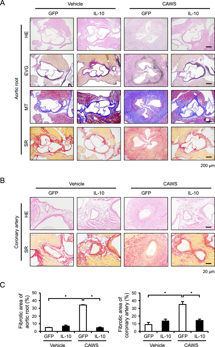
The AAV-mediated induction of IL-10 reduces CAWS-induced vascular inflammation and fibrosis Mice were treated intraperitoneally with CAWS or vehicle, 2 weeks after the injection of AAV-GFP (control) or AAV-IL-10. Heart sections were obtained at day 28 after CAWS or vehicle treatment and stained with HE, EVG, MT, and SR. (A,B) Representative images of the aortic root and coronary artery are shown. (C) The fibrotic area of the aortic root and coronary artery was quantified (n = 3–4). Data are expressed as the mean ± SEM. *p < 0.05.

