Figure 2.
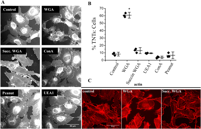
The sialic acid specificity of WGA induces endothelial TNT formation and F-actin rearrangements. (A) Examples of cell masked stained HUVECs following 30 mins incubation with the stated lectins (10 μg/ml). Scale bar is 50 μm. (B) Mean data and analysis of the effect of lectins on the percentage of TNTs to cells (n = 3, control N = 28, WGA N = 29, Succin WGA N = 30, UEA1 N = 30, ConA N = 29, peanut N = 30). Data are represented as mean ± SD, *P < 0.05. (C) Phalloidin staining (red) indicates F-actin redistribution in HUVECs treated with 10 μg/ml WGA for 30 mins as compared to control cells or cells treated with 10 μg/ml succinylated WGA. Scale bar is 50 μm.

