Fig. 1.
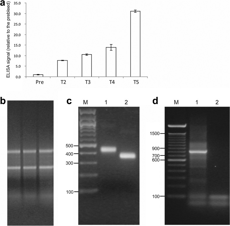
Successful immunization of a rabbit with C. jejuni WCA and amplification of scFv cDNA fragments from spleen of the rabbit. a ELISA signal against C. jejuni cells, using pre-bleed and antisera from the rabbit after two (T2), three (T3), four (T4), and five (T5) immunizations. Each column represents the mean fold difference in ELISA signal from pre-bleed. The error bars represent standard deviation of the signals in the triplicate wells for each antibody. b Agarose gel image of the three total RNA samples purified from the spleen of the rabbit immunized with C. jejuni WCA. The sharp rRNA bands show that the RNA samples had no degradation. c Agarose gel image of purified cDNA fragments for VH and VL chains amplified from immune rabbit spleen. PCR products using nine pairs of VK and 1 pair of Vλ primers were pooled together to purify the VL fragment. PCR products using four pairs of VH primers were pooled together to purify the VH fragment. Lane M = 100 bp ladder; bp size of some bands is shown on the left of the image. Lane 1 = ~ 450 bp VH cDNA, lane 2 = ~ 350 bp VL cDNA. d Agarose gel image of purified full-length scFv cDNA fragment after merging the VL and VH fragments by overlap extension PCR. Lane M = 100 bp ladder. Lane 1 = ~ 800 bp scFv cDNA fragment, lane 2 = non-template PCR control

