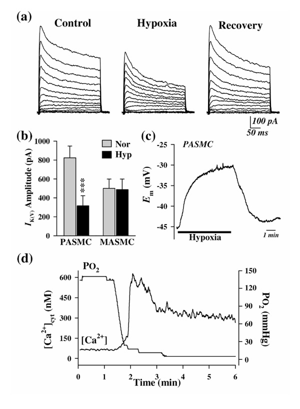
Figure 1.

Effects of acute hypoxia on whole-cell IK(V), Em, and [Ca2+]cyt in PASMCs. (a) Representative families of currents elicited by depolarizing the cell to test potentials ranging from -50 to +80 mV in 10-mV increments (holding potential -70 mV); these were recorded before, during, and after reduction of partial oxygen tension (PO2)in the extracellular solution from 149 to 8 mmHg. (b) Summarized data showing the effect of acute hypoxia (Hyp) on IK(V), elicited by a test pulse of +60 mV, in PASMCs and mesenteric artery smooth muscle cells (MASMCs). Data are expressed as mean ± standard error. ***P < 0.001 versus normoxia (Nor). (c) Change in Em recorded in a PASMC on switching from normoxic to hypoxic bath solutions. (d) [Ca2+]cyt measured (using Fura-2) in a peripheral cytoplasmic region of a PASMC on switching from normoxic to hypoxic bath solutions. The PO2 level was measured using an oxygen meter positioned in the tissue chamber. Modified with permission from Yuan et al [19**].
