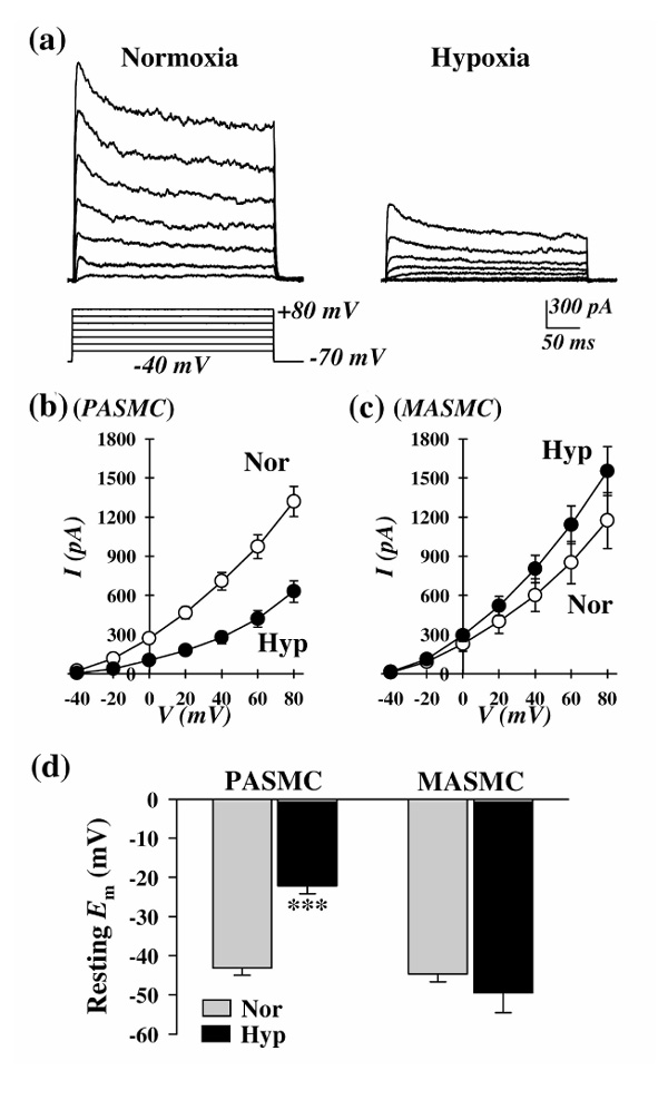
Figure 3.

Effects of prolonged hypoxia (3 days) on whole-cell IK(V) and resting Emin PASMCs and mesenteric artery smooth muscle cells (MASMCs). (a) Representative families of currents, elicited by depolarizing the cell to a series of test potentials ranging from -40 to +80 mV (holding potential -70mV) in PASMCs incubated in normoxia or hypoxia (25-35 mmHg). (b) and (c) Current-voltage relationship (I-V) curves (mean ± standard error) of whole-cell IK(V) in (b) PASMCs and (c)MASMCs incubated in normoxia (white circle) or hypoxia (black circle). (d) Summarized data (mean ± standard error) showing resting Em in PASMCs and MASMCs incubated in normoxia (Nor) or hypoxia (Hyp, partial oxygen tension 25-35 mmHg, for 60 h). ***P < 0.001 versus Nor.
