Figure 2.
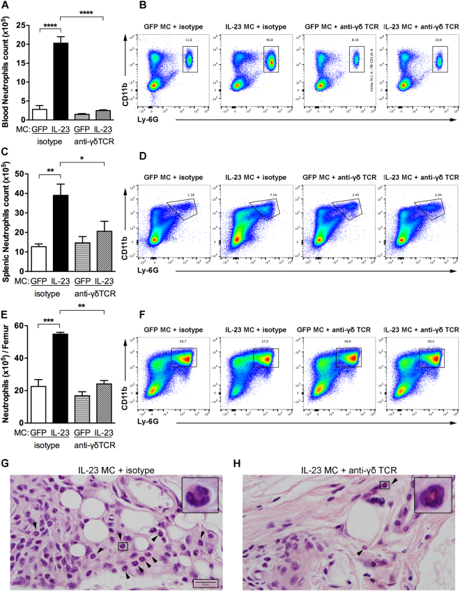
Anti-γδ TCR treatment inhibits the expansion of neutrophils. (A) Quantification of CD11b+ Ly-6G+ blood neutrophils in B10.RIII mice injected with GFP or IL-23 MC and treated with anti-γδ TCR or isotype mAb determined by flow cytometry. (B) Representative flow cytometry plots showing the gating strategy for evaluating the numbers of blood neutrophils. (C) Splenic CD11b+ Ly-6G+ neutrophil counts in B10.RIII mice injected with GFP or IL-23 MC and treated with anti-γδ TCR or isotype mAb determined by flow cytometry. (D) Gating strategy for evaluating the percentage and absolute numbers of neutrophils in mouse spleen. (E) Absolute numbers of CD11b+ Ly-6G+ neutrophils per femur. (F) Representative flow cytometry plots showing the gating strategy. Numbers depict the percentage of CD45+ leukocytes. (n = 4–5 per group). (G,H) Representative haematoxylin and eosin-stained sections of day-11 ankles histopathology from mice receiving isotype control or anti-γδ TCR mAb showing neutrophils in joint capsule imaged with 100 × oil-immersion objective lense. Arrows indicate polymorphonuclear neutrophils. Scale bars, 20 μm. Data were obtained from 3 independent experiments (n = 4–5 per group). All data are shown as mean ± SEM. *p < 0.05, **p < 0.01, ***p < 0.001, ****p < 0.0001. Statistical analysis was performed using one-way ANOVA with Sidak’s multiple comparisons test.

