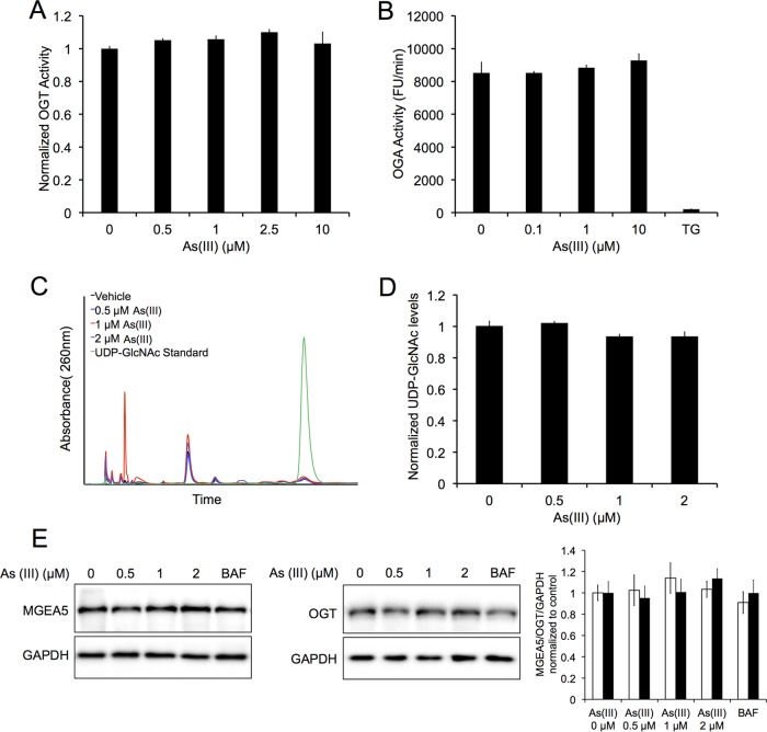
FIG 6.
OGT and OGA protein level and activity, as well as UDP-GlcNAc levels, are not affected by arsenic treatment. (A) Recombinant OGT was exposed to 0, 0.5, 1, 2.5, or 10 μM As(III) for 1 h and OGT activity was assessed based on the levels of released phosphate as indicated by changes in malachite green absorbance at 660 nm. The graph presents results normalized to the control. Data are means ± SEMs (n = 3). (B) Recombinant MGEA5 was exposed to a vehicle or 0.1, 1, or 10 μM As(III), and activity was assessed over a 5-min period by measuring the fluorescence of the fluorogenic substrate 4-methylumbelliferyl N-acetyl-β-d-glucosaminide (4-MU). A 10 μM concentration of thiamet G (TG) was used as a positive control. The graph presents MGEA5 activity (RFU/min) normalized to the control. Data are means ± SEMs (n = 3). (C) HeLa cells were treated with 0, 0.5, 1, or 2 μM As(III) for 4 h. UDP-GlcNAc levels were then measured using high-performance liquid chromatography. The chromatogram indicates UDP-GlcNAc peak following As(III) treatment compared to a diluted UDP-GlcNAc standard. The graph presents UDP-GlcNAc levels normalized to the control. Data are means ± SEMs (n = 3). (D) HeLa cells were treated with 0, 0.5, 1, or 2 μM As(III), 100 nM BAF, or HBSS for 4 h. Immunoblot analysis of OGT or MGEA5 protein levels was conducted following treatment. GAPDH was used as an internal loading control. Data are means ± SEMs (n = 3).

