
FIG 1.
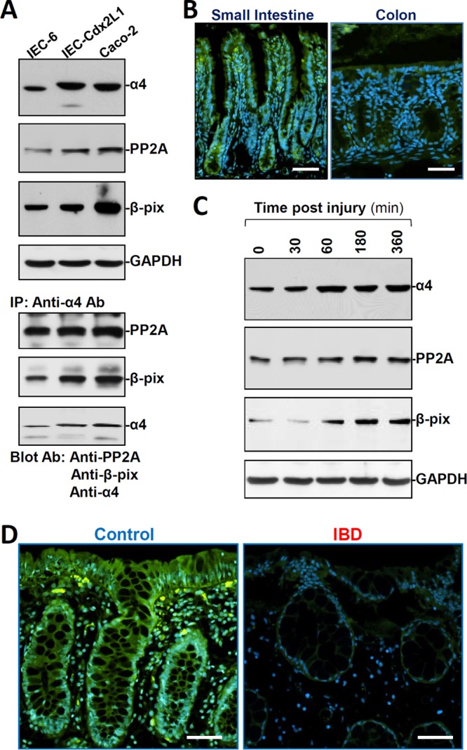
Expression levels of α4 in the intestinal epithelium with or without pathological stress. (A) Representative immunoblots of α4, PP2A, and β-PIX proteins (top) and their interaction (bottom) in different lines of cultured IECs. In studies examining protein-protein interactions, whole-cell lysates (400 μg) were immunoprecipitated by using an antibody (Ab) that recognizes α4, and the levels of PP2A, β-PIX, and α4 in the products of the IP reactions were examined by Western blotting. (B) Immunostaining of α4 (green) in mouse mucosal tissues from the small intestine and colon. Scale bars, 50 μm. (C) Changes in the levels of α4, PP2A, and β-PIX after wounding in cultured differentiated IEC-Cdx2L1 cells. After the cells were grown to confluence, epithelial repair was induced by removing part of the monolayer. The levels of α4, PP2A, and β-PIX were examined at different times after wounding. (D) Immunostaining of α4 in human intestinal mucosal tissues from control individuals (without mucosal erosions/inflammation) and from patients with IBD. The experiments were repeated in samples obtained from four patients with IBD or control individuals and showed similar results. Scale bars, 50 μm.