
FIG 2.
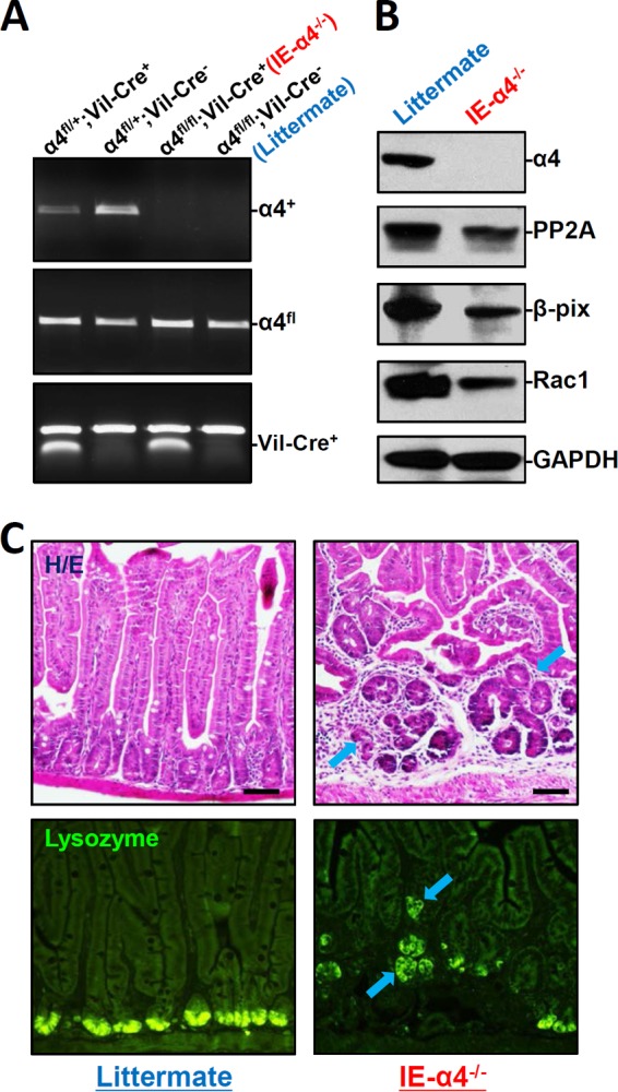
α4 deletion in IECs disrupts mucosal maturation in the small intestine. (A) PCR analysis of genomic DNA from the small intestinal mucosa indicating floxed, α4 deletion, and Vil-Cre bands in mice with different genotypes. (B) Immunoblots of α4, PP2A, Rac1, and β-PIX proteins in the small intestinal mucosa obtained from controls (littermate) and intestinal epithelial tissue-specific α4 knockout (IE-α4−/−) mice. (C) Photomicrographs of hematoxylin and eosin (H/E) (top) and immunostaining of lysozyme (bottom), shown as in green, in the small intestine. Scale bars, 50 μm.