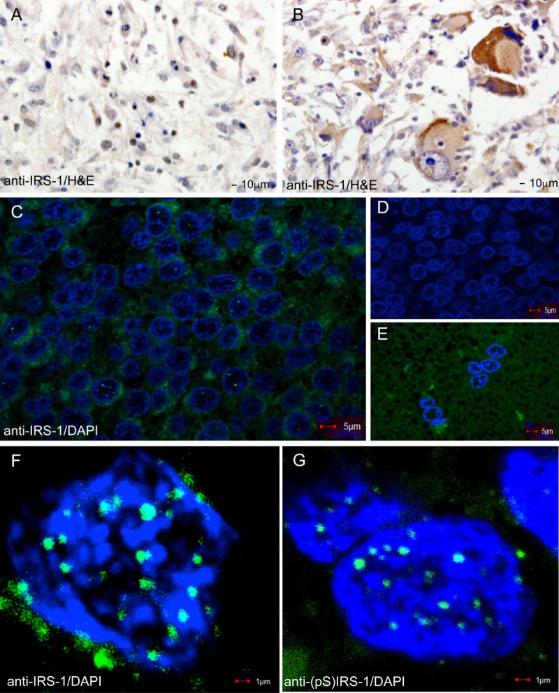
FIG 1.

Immunohistochemical detection of IRS-1. IRS-1 was detected in formalin-fixed paraffin-embedded sections of glioblastoma tissues (GL803a tissue array; USBiomax, Inc.) by using anti-IRS-1 rabbit polyclonal (Millipore) and anti-rabbit HRP-conjugated secondary antibodies. (A and B) Two glioblastoma biopsy specimens in which IRS-1 is localized in either the nuclei of some tumor cells (case C2 from Table 1) (A) or the cytoplasm (case A5 from Table 1) (B). (C, F, and G) Examples of low-magnification (C) and high-magnification (F and G) confocal images of IRS-1 nuclear structures detected in a restricted number of cells from glioblastoma biopsy specimens. IRS-1 nuclear structures were detected with anti-IRS-1 rabbit polyclonal antibody (catalog no. 06-248; Millipore) and FITC-conjugated anti-rabbit secondary antibodies. Nuclei were counterstained with DAPI (blue fluorescence). (C) Selected area adjacent to the tumor-infiltrating margin with a high number of tumor cells positive for IRS-1 nuclear structures (15 out of a total of 121 cells show IRS-1 nuclear structures). The percentage of tumor cells positive for IRS-1 nuclear structures was evaluated by using high-magnification confocal imaging (original magnification, ×100). At least 100 randomly selected fields per biopsy specimen were examined for 10 different glioblastoma biopsy specimens. (D) The same glioblastoma multiforme biopsy specimen immunolabeled with an irrelevant antibody (anti-BrdU primary antibody and FITC-conjugated secondary antibody). (E) Unaffected brain area, adjacent to the tumor depicted in panel C, immunolabeled with anti-IRS-1 rabbit polyclonal antibody.
