FIG 3.
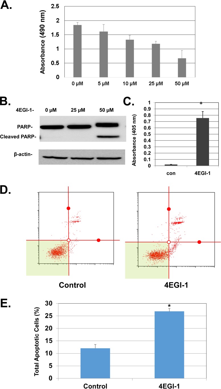
(A) 4EGI-1 inhibits the viability of LNCaP cells in a dose-dependent manner. Subconfluent cells were treated with 4EGI-1 at the indicated concentrations for 48 h. After treatment, an MTT assay was performed as described in Materials and Methods. The results are averages ± the SEM from three repeats. (B) 4EGI-1 induces PARP cleavage in LNCaP cells. Subconfluent cells were treated with the indicated concentrations of 4EGI-1 for 24 h. After treatment, both floating and attached cells were harvested and lysed. Equal amounts of protein were then subjected to SDS-PAGE and transferred to PVDF membranes. PARP, cleaved PARP, and β-actin were detected by immunoblotting. The results are representative of three repeats. (C) 4EGI-1 causes DNA fragmentation in LNCaP cells. Subconfluent cells were treated with 50 μM 4EGI-1 for 24 h. Both floating and attached cells were harvested and lysed after 4EGI-1 treatment. Fragmented DNA was measured by an ELISA cell death assay as described in Materials and Methods. The results are averages ± the SEM from three repeats (*, P < 0.05). (D and E) 4EGI-1 induces apoptosis in LNCaP cells: the cell apoptosis assay was performed using the Muse annexin V and dead cell assay kit (Millipore) according to the manufacturer's protocol. Briefly, LNCaP cells were grown to subconfluence and then treated with 50 μM 4EGI-1 for 24 h. After the treatment, both floating and attached cells were harvested and then labeled with Muse annexin V and dead cell reagent as described in Materials and Methods. After incubation, the tubes were read individually (as shown in panel D) using the Muse cell analyzer (Millipore). The averages of total apoptotic cells (early apoptotic and late apoptotic) ± the SEM from three individual experiments are presented in panel E (P < 0.05).

