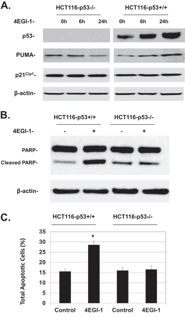
FIG 5.

(A) 4EGI-1 induces p53 and PUMA but not p21Cip1 in HCT116-p53+/+ cells. HCT116-p53+/+ and HCT116-p53−/− cells were treated with 50 μM 4EGI-1 for the indicated time points. After treatment, the cells were lysed. p53, PUMA, p21Cip1, and β-actin were detected following SDS-PAGE and Western blotting. (B) 4EGI-1 induces PARP cleavage in HCT116-p53+/+ cells. Subconfluent HCT116-p53+/+ and HCT116-p53−/− cells were treated with 50 μM 4EGI-1 for 24 h. After treatment, both floating and attached cells were harvested and lysed. Equal amounts of protein were then subjected to SDS-PAGE and transferred to PVDF membranes. PARP, cleaved PARP, and β-actin were detected by immunoblotting. Results in panels A and B are representative of three repeats. (C) 4EGI-1 induces apoptosis in HCT116-p53+/+ cells. HCT116-p53+/+ and HCT116-p53−/− cells were grown to subconfluence and then treated with 50 μM 4EGI-1 for 24 h. Following the treatment, both floating and attached cells were harvested and then labeled with Muse annexin V and dead cell reagent as described in Materials and Methods. After incubation, the tubes were read using a Muse cell analyzer (Millipore). The averages of total apoptotic cells (early apoptotic and late apoptotic) ± the SEM from three individual experiments are presented (P < 0.05).
