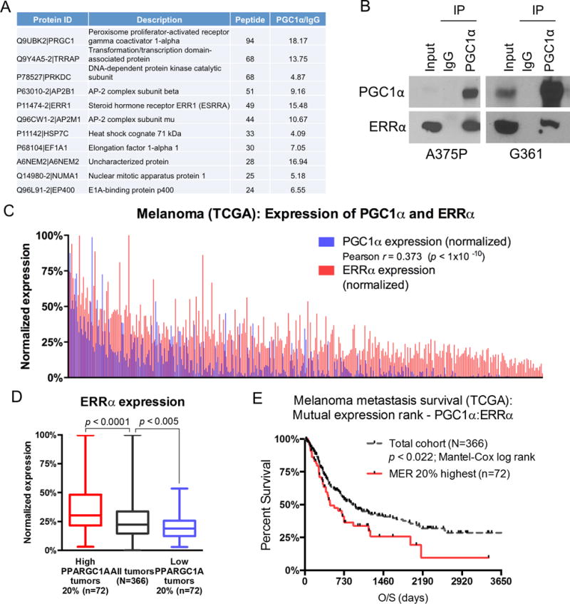
Figure 1. ERRα is associated with PGC1α in melanoma cells.

A. A list of the most abundant proteins co-immunoprecipitated with Flag/HA-tagged PGC1α in A375P melanoma cells as identified by mass spectrometry. B. Endogenous PGC1α is interacting with ERRα in PGC1α-positive melanoma cell lines A375P and G361. Immunoprecipition of PGC1α was followed by immunoblotting using the indicated antibodies. C. Pearson-based correlation between PGC1α and ERRα expression levels across metastatic melanoma samples within TCGA (N=366). D. Based on 2-sample, 2-sided t-test statistics, the 20 percentile highest and lowest PGC1a expression levels associate with highest and lowest ERRα expression levels, respectively. E. Based on Mantel-Cox log rank test, the top 20 percentile mutual expression rank (MER) of PGC1α:ERRα segregates poorer overall survival relative to the metastatic melanoma cohort average (p < 0.022).
