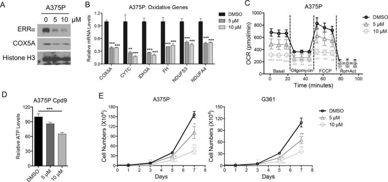
Figure 4. Inhibition of ERRα activity phenocopies ERRα depletion in melanoma cells.

A. Immunoblotting of melanoma cells upon treatment with ERRα antagonist Cpd29. B. Expression of oxidative genes at the mRNA level in melanoma cells upon ERRα antagonist Cpd29. C. Mitochondrial activity of melanoma cells treated with ERRα antagonist Cpd29 as measured by seahorse flux assay. D. Intracellular ATP levels in cells treated with Cpd29. E. Growth curve of various melanoma cell lines treated with Cpd29. *P < 0.05, **P < 0.01, ***P < 0.001 as determined by t-test (B, E) or one-way ANOVA (C, D).
