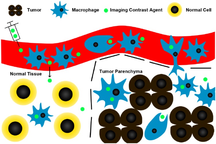
Figure 1.
Contrast agents, such as iron oxide nanoparticles, are preferentially phagocytosed by monocytes and macrophages, and can be used to label TAMs. Dotted lines represent the tumor boundaries. Contrast agents are injected intravenously where they are phagocytosed by circulating monocytes. Some of these monocytes that have phagocytosed the contrast agent migrate into the tumor and differentiate into macrophages. As tumors often have increased vascular permeability, contrast agents could also leak into the tumor and be picked up by macrophages. Post-contrast images are usually acquired at least 24 hours after administration of contrast, to allow time for phagocytosis/cell migration, and wash out of non-phagocytosed contrast agent. Although macrophages in normal tissue may still take up contrast agents, there will be fewer of them, and the normal vascular permeability may decrease contrast leakage into normal tissue.

