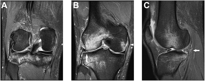
Figure 2.
(A, B) Coronal proton density–weighted fat-saturated images and (C) fat-suppressed T2-weighted image illustrating bone bruising in both the medial and lateral compartments with a grade 2 injury of the medial collateral ligament (white triangle) with a lateral meniscal tear (solid arrow) in the posterior horn and body.

