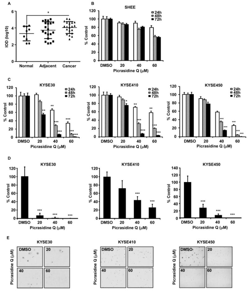
Figure 3.

PQ suppressed the growth of ESCC cells. (A) Expression of FGFR2 on ESCC tissues. (B) Toxicity of PQ on normal esophageal cells. (C) Effects of PQ on the proliferation of KYSE30, KYSE410 and KYSE450 ESCC cells were assessed at 24, 48 and 72 h by MTT assay. The asterisk indicates a significant decrease in proliferation compared with vehicle control (**P < 0.01, ***P<0.001). (D) Effects of PQ on anchorage-independent growth of KYSE30, KYSE410 and KYSE450 ESCC cells were evaluated. The asterisks (***P < 0.001) indicate a significant decrease in colony formation with PQ treatment compared with untreated control. (E) Reflected pictures of anchorage-independent cell growth results.
