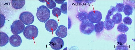
Fig. 1.

Myeloblasts in bone marrow smears from mice in the WEHI-3 and WEHI-3 + P. yoelli (Py) groups. Mice were i.p. injected with WEHI-3 cells. After 3 weeks, the bone marrow was flushed from the femurs of the sacrificed mice and smeared. Leukocyte classification based on cell morphology was performed by Wright’s staining of the bone marrow smears. The arrows (↓) indicate neoplasm cells (myeloblast), which contain large, irregular nuclei accompanied by prominent nucleoli and abundant light eosinophilic cytoplasm
