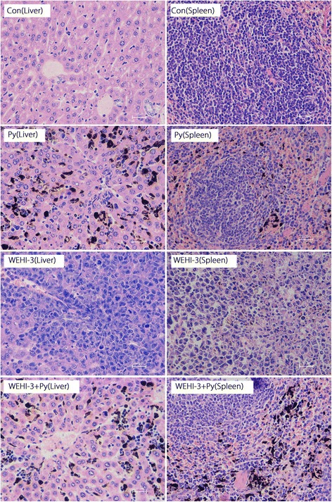
Fig. 3.

Histopathological examination of liver and spleen tissues from each group. The isolated spleen and liver samples were fixed in 4% formaldehyde, embedded in paraffin and sectioned. The sections (5 μm) were stained with H&E and evaluated under a microscope at 400 × magnification. The Con group shows normal structures and no infiltrated neoplasm cells in the liver and spleen. The P. yoelli (Py) group shows normal structures and no infiltrated neoplasm cells but shows deposition of malaria pigments in the liver and spleen. The WEHI-3 group shows abundant infiltrated neoplasm cells in the liver and spleen. The WEHI-3 + Py group shows markedly reduced neoplasm cell infiltration in the liver and spleen
