
Fig. 5.
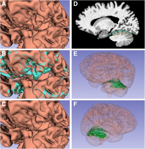
Manual smoothing of cortical surface and exclusion of the tentorium
Panel a: White and gray matter imported from SPM included protrusions and nodules that required editing. These bumps likely represent dura, cortical veins and arteries, and sinuses. Panel b: Protrusions, manually highlighted in turquoise, are removed to produce a smooth cortical surface shown in Panel c. Panel d: Representative sagittal slice showing the manual segmentation of the tentorium, which is subtracted from the final brain segmentation. 3D rending of the tentorium from the left (Panel e) and right (Panel f), with the rest of the brain transparent