
Fig. 8.
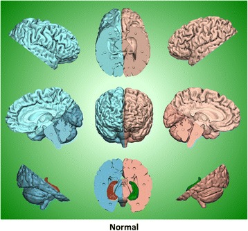
Normal brain STL files. The final STL files of the normal brain are displayed showing the cutting planes, sectional breakdown and magnet holes. The following STL images in Figs. 7 and 8 and the photographs in Figs. 9 and 10 are all displayed at the same scale, so the contrast in size between the normal and severe Alzheimer’s brains pictured here is representative of the disparity displayed by the physical models themselves