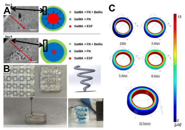
Figure 3. 3D printed placenta mimetic.
A) Illustration and corresponding micrographs demonstrating the invasion of cells printed onto the outer periphery of the mimetic with an EGF source in the middle of the scaffold at day 2 and day 8 of the study. B) This work demonstrates the ability to fabricate complex scaffold using multiple soft hydrogel materials to create defined regions with different components. C) COMSOL modeling of the EGF gradient established over the time course of the experiment throughout the hydrogel. Models such as this can be readily developed using the material properties and CAD models to theoretically determine the concentration of molecules as a function of time within 3D printed scaffolds. Reproduced and adapted with permission.[41] Copyright 2017, American Chemistry Society.

