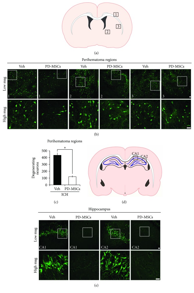
Figure 3.
Human placenta-derived mesenchymal stem cells (PD-MSCs) reduced neuronal death in the brains of rats subjected to intracranial hemorrhage (ICH). (a) The location of core hemorrhagic regions at 0.2 mm from the bregma. (b) Fluorescence images reveal the degenerating neurons in the perihematomal region at 24 hours after ICH. Degenerating neurons are detected by Fluoro-Jade B (FJB) staining (green). Scale bar = 20 μm. (c) The bar graphs represent the count of FJB-positive neurons in the perihematomal region from the vehicle-treated and the PD-MSC-treated groups at 24 hours after ICH induction. Data are mean + SEM; n = 7 − 8 from each group (ICH-Veh, n = 7; ICH-MSC, n = 8), ∗p < 0.05. (d) The location of hippocampal regions at −3.6 mm from the bregma. (e) Fluorescence images reveal the degenerating neurons only in the hippocampal CA1 and CA2 region of the vehicle-treated group at 24 hours after ICH. Scale bar = 20 μm.

