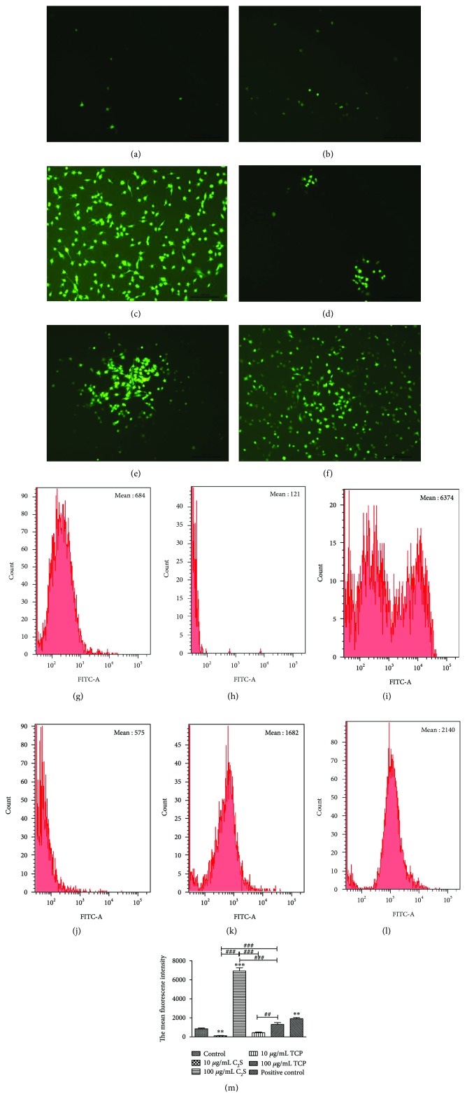
Figure 5.
(a), (b), (c), (d), (e), and (f) were captured using an inverted fluorescence microscope, while (g), (h), (i), (j), (k), and (l) were acquired using FCM, which also produced the mean fluorescence intensity of the groups. (a and g) Control group, (b and h) 10 μg/mL C2S particle group, (c and i) 100 μg/mL C2S particle group, (d and j) 10 μg/mL TCP particle group, (e and k) 100 μg/mL TCP particle group, and (f and l) positive control group. (m) A cartogram of the mean fluorescence intensity. The 10 μg/mL C2S (compared with control group, P < 0.01) and TCP particle groups showed no obvious ROS and had even lower levels than in the control group. However, when the concentration was brought up to 100 μg/mL, a large amount of ROS was produced. Moreover, the 100 μg/mL C2S particle group generated more ROS than the 10 μg/mL (P < 0.001) and 100 μg/mL (P < 0.001) TCP particle groups as well as the 10 μg/mL C2S particle group, as shown above. Significant changes were observed between the 10 μg/mL and the 100 μg/mL TCP particle groups (P < 0.01) and the 10 μg/mL C2S particle group (P < 0.001). The RAW 264.7 cells produced obvious ROS when incubated with H2O2 (P < 0.01). ∗P < 0.05, ∗∗P < 0.01, and ∗∗∗P < 0.001 (experimental group versus control group). #P < 0.05, ##P < 0.01, and ###P < 0.001 (between the experimental groups).

