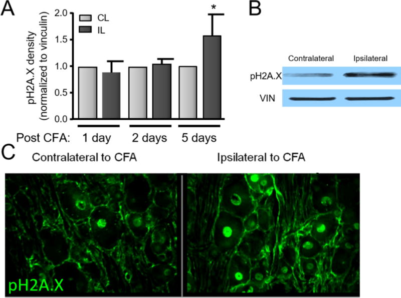
Figure 1.

DNA damage is enhanced in the lumbar DRG following hindpaw inflammation. A. Representative western blot of pH2A.X and vinculin (loading control) expression in contralateral and ipsilateral L4/L5 DRG 5 days following unilateral CFA injection into the rat hindpaw. B. Each column represents the mean ± SEM of the density of pH2A.X from 6 experiments normalized to the amount of vinculin. An asterisk indicates a statistically significant increase in the DRG ipsilateral to CFA injection compared to those contralateral to the injection using Student t-test. C. Photomicrographs (20X) of pH2A.X in L5 DRG from a rat 5 days after CFA injection. Green fluorescence indicates the immunoreactivity to pH2A.X.
