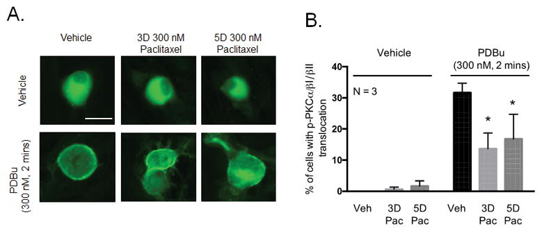
Figure 6.

Chronic treatment with paclitaxel decreases PDBu-stimulated membrane localization of PKC α, βI and βII in cultured sensory neurons. (A) Representative fluorescent images showing membrane localization of immunoreactive phosphorylated PKC α, βI and βII in neurons treated with paclitaxel (300 nM, 3 days or 5 days) following stimulation with PDBu (300 nM, 2 minutes). (B) Graphical analysis showing the number of neuronal cell bodies demonstrating translocation of phosphorylated PKC α, βI and βII in paclitaxel-treated neurons (300 nM, 3 and 5 days) following stimulation with PDBu (300 nM, 2 minutes) expressed as % of phospho- PKC α/βI/II translocation. An * indicates a significant decrease in the number of cells with PKC α, βI and βII translocation. Significance was determined using a two-way ANOVA with Dunnett’s post-hoc test (p < 0.05, N = 3). Scale bar = 20 μm. Veh - Vehicle; Pac –Paclitaxel, 3D – 3 days, 5D – 5 days.
