Figure 7.

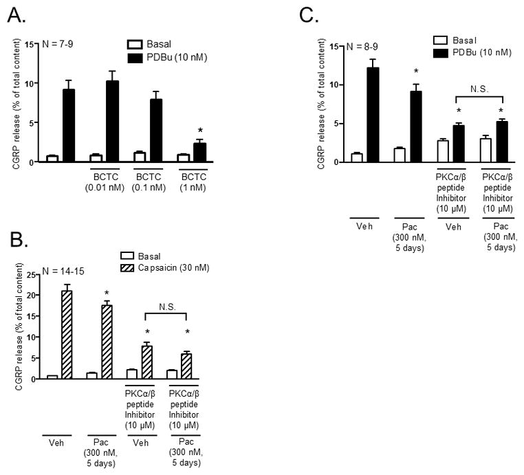
Inhibition of PKCα and PKCβI/II activity mediates the decrease in capsaicin-stimulated CGRP release following chronic exposure to paclitaxel in cultured sensory neurons. Each column represents the mean ± SEM of basal (white columns), capsaicin-stimulated (striped columns) or PDBu-stimulated (black columns) CGRP release expressed as % of total content. (A) Naïve cultures were pre-treated with the TRPV1 antagonist, BCTC, for 10 minutes prior to stimulation with capsaicin (30 nM). An * indicates a significant decrease in capsaicin-stimulated release in neurons pre-treated with BCTC compared to control neurons (p < 0.05, N = 7–9). Cultures were exposed to 300 nM paclitaxel for 3 days and pre-treated with a myristolyated PKCα/β peptide inhibitor (10 μM) for 10 minutes prior to stimulation with (B) 30 nM capsaicin or (C) 10 nM PDBu. An * indicates a decrease in (B) capsaicin-stimulated and (C) PDBu-stimulated release in paclitaxel-only treated neurons, vehicle- and paclitaxel treated neurons pre-treated with the myristolyated PKCα/β peptide inhibitor compared to vehicle-only treated neurons. Significance was determined using a two-way ANOVA with Tukey’s post-hoc test (p < 0.05, N = 8–15). N.S. – not significant.
