Figure 2.
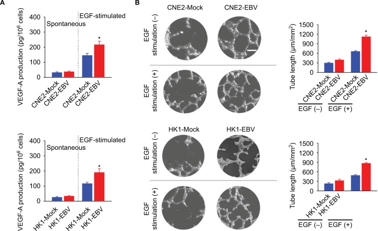
EBV infection promotes EGF-stimulated VEGF production and endothelial tube formation.
Notes: (A) VEGF-A production was determined in the mock-controlled and the EBV-infected CNE2 or HK1 cell-conditioned medium. The amount of VEGF released from the serum-starved cells in the absence or presence of extracellular EGF stimulation was determined by ELISA. (B) HUVECs were incubated with the cell-conditioned medium as indicated. Representative photographs of HUVEC tube formation were captured at 6 hours after cell seeding; scale bar =100 μm. The tube formation was quantitatively evaluated by calculating the tube length per standard area in each well (right panel). The data are representative of three independent experiments and are presented as the mean ± SEM (*P<0.05, Student’s t-test).
Abbreviations: EBV, Epstein–Barr virus; EGF, epidermal growth factor; ELISA, enzyme-linked immunosorbent assay; HUVECs, human umbilical vein endothelial cells.

