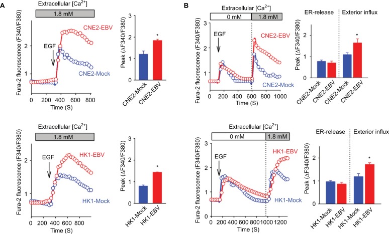
Figure 3.
EBV infection amplifies EGF-stimulated Ca2+ signaling via SOCE.
Notes: (A) EGF-evoked Ca2+ transient responses were measured in the fura-2-loaded cells. The dynamic cytosolic Ca2+ level [Ca2+]cyt was measured as the fluorescence ratio (F340/F380) of fura-2. Each trace represents the average data from at least 20 individual cells. The intensities of Ca2+ responses were quantitatively evaluated by calculating the average peak from the baseline in each right panel. (B) EGF-induced Ca2+ release from intracellular Ca2+ stores (ER) and the following Ca2+ influx via membrane channels were measured in the absence and presence of extracellular Ca2+, successively. The data are representative of three independent experiments and are presented as the mean ± SEM (*P<0.05, Student’s t-test).
Abbreviations: EBV, Epstein–Barr virus; EGF, epidermal growth factor; ER, endoplasmic reticulum.

