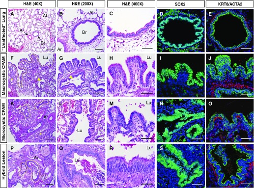
Figure 2.
Congenital lung lesion cysts resemble disordered airway structures. (A–C) Histological analysis of unaffected lung tissue. (D and E) SOX2 and KRT8 (green)/ACTA2 (red) expression in unaffected lung tissue, demonstrating a normal bronchovascular bundle surrounded by alveolar tissue. (F–H, K–M, and P–R) Histological analysis of macrocystic (F–H), microcystic (K–M), and hybrid lesion (P–R) tissue. (I, J, N, O, S, and T) SOX2 and KRT8 (green)/ACTA2 (red) expression in macrocystic (I and J), microcystic (N and O), and hybrid lesion (S and T) tissue. A large cystic structure (lumen outlined by dashed yellow line in F) is evident in a macrocystic congenital pulmonary airway malformation lesion, which is lined by papillary epithelial structures varying in thickness from columnar to pseudostratified (F–J). Smaller cysts (indicated by yellow arrowheads) and compressed alveolar tissue (indicated by yellow asterisks) are also evident. Smaller cysts are highlighted with dashed yellow lines in K and P. ACTA2 = smooth muscle α-actin; Ar = artery; Al = alveolar region; Br = bronchiole; CPAM = congenital pulmonary airway malformation; H&E = hematoxylin and eosin; KRT8 = keratin, type II cytoskeletal 8; Lu = lumen; SOX2 = SRY (sex-determining region Y)-related HMG box protein 2. Scale bars: (A, F, K, and P) 500 μm; (B, G, L, and Q) 100 μm; all others, 50 μm.

