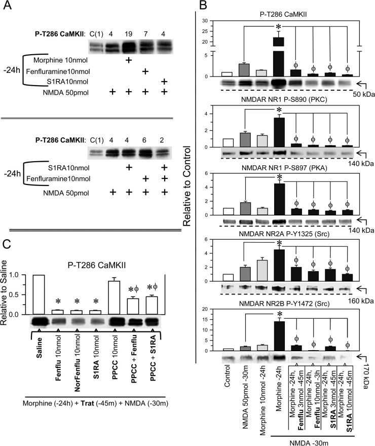
Figure 4. Effect of fenfluramine and S1RA on the overactivity of NMDARs promoted by NMDA in morphine-primed mice.
(A) Morphine-primed mice exhibited a high level of CaMKII auto phosphorylation in response to NMDA. This effect was not evoked by fenfluramine or S1RA, which instead diminished P-T286 CaMKII levels in those morphine and NMDA-treated mice. The assay was repeated at least twice. The immunosignals are expressed relative to the control (C, value of 1), which received saline instead of the treatments indicated. (B) Effect of the different treatments on NMDAR phosphorylation and on P-T286 CaMKII. All substances were given icv, and the mice were killed at the post-drug intervals indicated, e.g., 30 min after NMDA and 24 h after morphine. The combined treatments started with morphine, and 24 h later, fenfluramine or S1RA was administered 45 m before NMDA. * Significant difference with respect to the group that received only morphine; ϕ significant difference respect to the morphine-primed group that received only NMDA 24 h later; ANOVA, Dunnett multiple comparisons vs control group, p < 0.05. (C) Effect of the σ1R agonist PPCC on the diminished effect of fenfluramine and S1RA on CaMKII hyper phosphorylation evoked by NMDA. The mice were primed with morphine, and 24 h later, the effects of the different drugs on P-T286 CaMKII were evaluated. *Significant difference with respect to the morphine-primed group that received saline instead of the drug and NMDA 24 h later; ϕ significant difference with respect to the group that received fenfluramine or S1RA but not PPCC; ANOVA, Dunnett multiple comparisons vs control group, p < 0.05. (A–C) Each bar represents the mean ± SEM of 6 mice. Details as in Figure 3 and Supplementary Figure 3.

