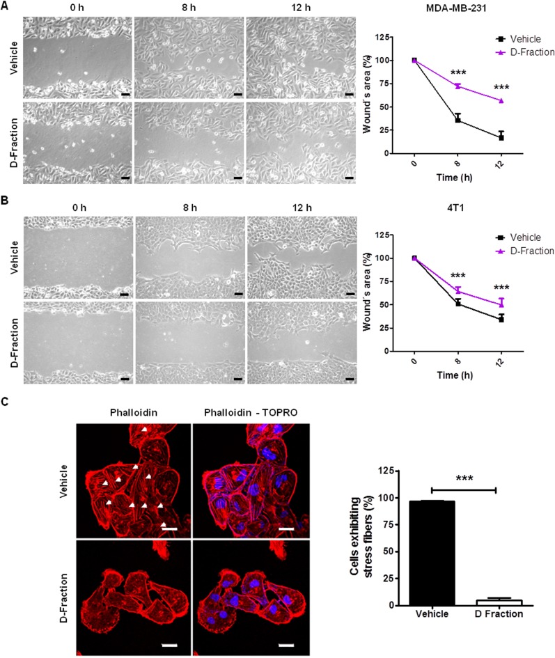
Figure 2. Maitake D-Fraction decreases migration of TNBC MDA-MB-231 and 4T1 cells and reduces the presence of stress fibers.
Representative phase-contrast pictures of the wound healing assay of (A) MDA-MB-231 cells and (B) 4T1 cells under treatment with D-Fraction (IC50) or vehicle. Magnification: 200×. The graphs represent the mean percentage (±SD) of uncovered wound area taking the value at 0 h as 100% of one representative experiment. Two-way ANOVA and Bonferroni post tests were performed. (C) Representative fluorescence images of MDA-MB-231 cells stained with rhodamine-conjugated phalloidin (F-actin) and TOPRO (nuclei) after treatment with D-Fraction (IC50, 12 h) or vehicle. Arrows indicate stress fibers. The graph represents the mean percentage (±SD) of cells exhibiting stress fibers after treatment of one representative experiment. Magnification: 630×, scale bars represent 20 μm. Student´s t test was applied. ***p < 0.001.

