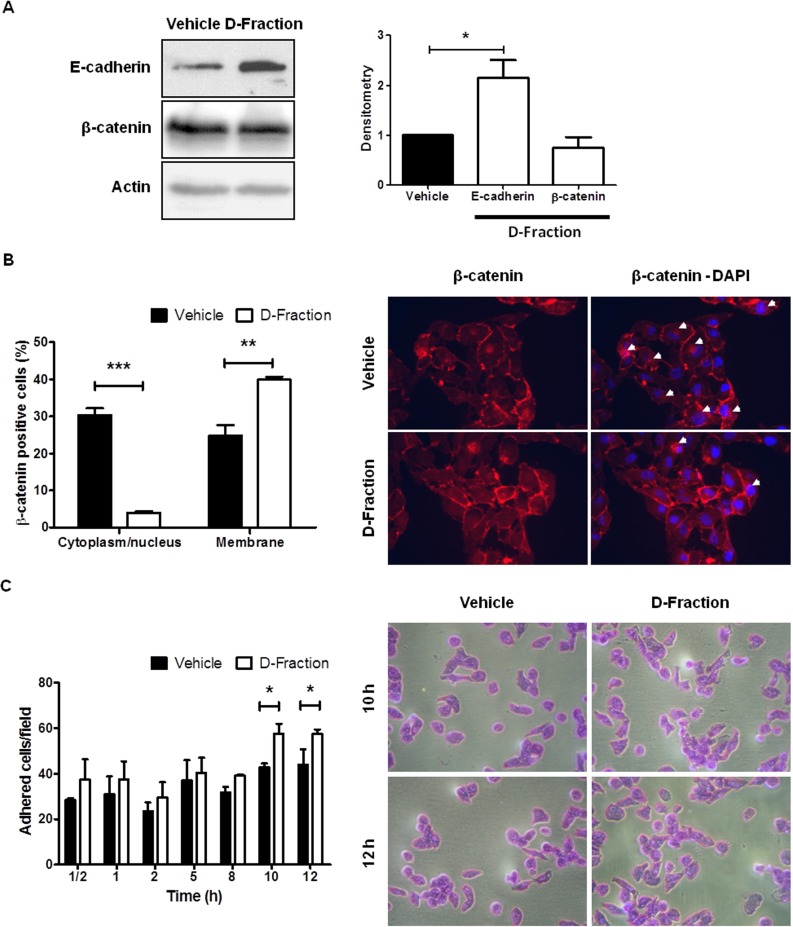
Figure 4. Maitake D-Fraction promotes the intercellular adhesion and cell-substrate adhesion in TNBC MDA-MB-231 cells.
(A) WB analysis for E-cadherin and β-catenin protein in cells treatment with D-Fraction (IC50, 12 h) or vehicle. Representative blots of at least two independent experiments and densitometry, mean ± SD. Student´s t test. (B) IF for β-catenin after treatment with D-Fraction (IC50, 12 h) or vehicle. The graph represents the proportion of cells (mean ± SD) with expression of β-catenin in cytoplasm/nucleus or membrane after treatment. Representative fluorescence images; arrows indicate the cytoplasm localization of β-catenin. Magnification: 400x. Student's t test. (C) Cell adhesion assay after treatment with D-Fraction (IC50, 12 h) or vehicle. The graph represents the number of adhered cells (mean ± SD) after treatment of one representative experiment. Representative pictures of cell adhesion assay. Magnification: 400x. Two-way ANOVA and Bonferroni post tests. *p < 0.05, **p < 0.01, ***p < 0.001.

