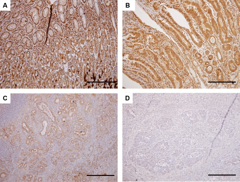
Figure 3. Immunohistochemical analysis for HHLA2 protein expression in primary tumor specimens.
On the basis of staining intensity, the status of HHLA2 expression was classified into the following three groups: high, intermediate, and low immunoreactions. (A) HHLA2 protein expression in normal epithelial cells. (B) Tumor cells with high expression of HHLA2. (C) Tumor cells with intermediate expression of HHLA2. (D) Tumor cells with low expression of HHLA2. Scale bars indicate 200 μm (Original magnification ×200).

