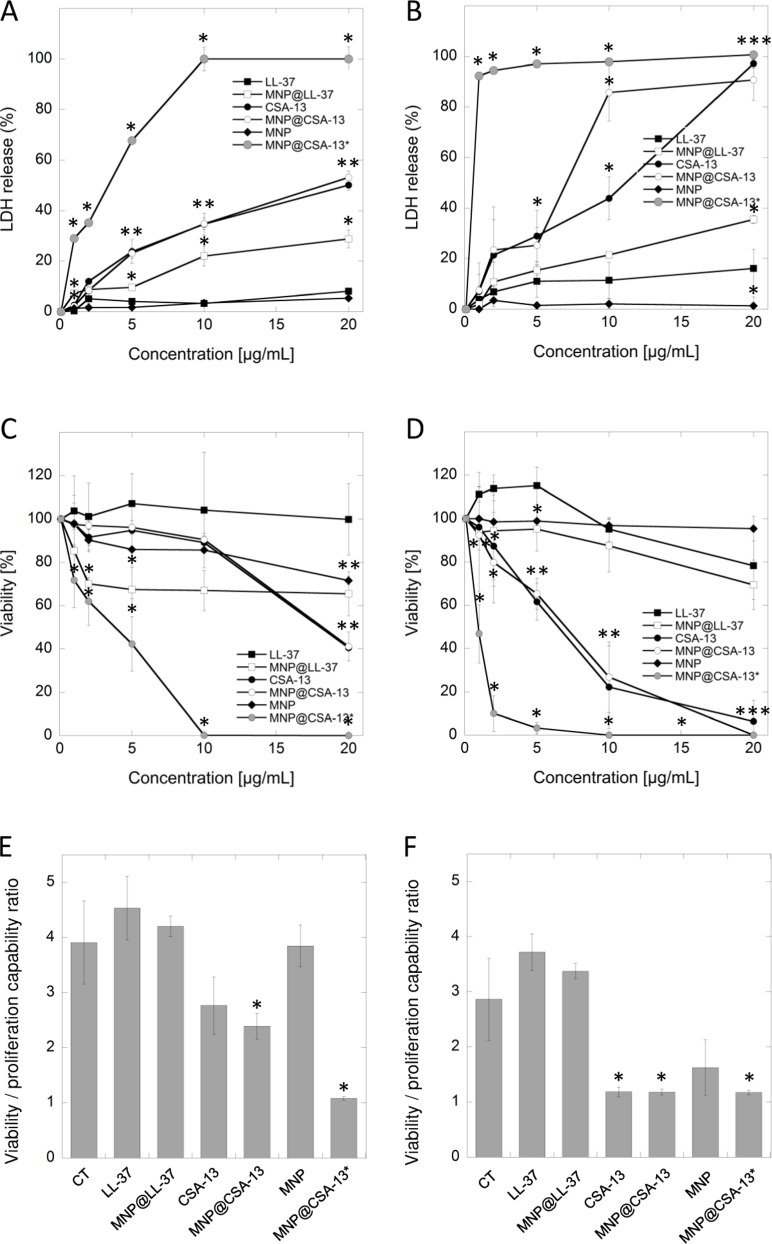
Figure 2.
Cytotoxic activity of cationic lipids and their magnetic derivatives against breast cancer MCF-7 (A, C, E) and MDA-MB-231 (B, D, F) cell lines. Increased LDH release from cancer cells (panels A and B) and the decrease of cell viability assessed by MTT assay (panels C and D) after 24 h incubation of cancer cells with varied concentrations of LL-37 peptide (black square), MNP@LL-37 (white square), CSA-13 (black circle), MNP@CSA-13 (white circle) and uncoated MNPs (black diamond). MNP@CSA-13* (grey circle) indicate the alternations in cell viability after treatment with MNP@CSA-13 in doses corresponding to the amounts of CSA-13 immobilized in the nanosystem (assuming the immobilization efficiency of approx. 14%). Panels E and F demonstrate the proliferation of cancer cells treated for 24 hours with 20 µg/mL of tested agents when compared to untreated control (CT; black inverted triangle) estimated using resazurin-based fluorimetric method (panels E and F). Results represent mean ± SD from 3 to 6 independent experiments. * indicates statistical significance (p < 0.05) when compared to LL-37 activity (panels A–D) or untreated control (panels E and F).

