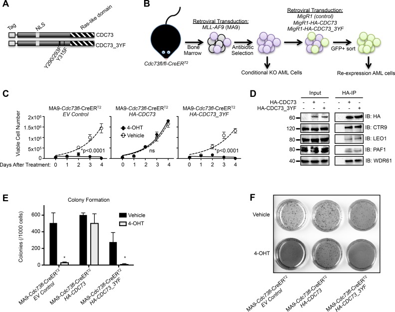
Figure 1. A CDC73 tyrosine mutant does not support AML cell growth.
(A) Schematic of HA-CDC73 showing known domains of CDC73 and the location of the three tyrosine mutations introduced to make CDC73_3YF. (B) Diagram representing the workflow for the generation of MLL-AF9 transformed CDC73fl/fl-CreERT2(MA9-Cdc73fl-CreERT2) CDC73 or CDC73_3YF re-expression cells, as well as MigR1 control cells (EV Control). (C) The indicated MLL-AF9 transformed CDC73fl/fl-CreERT2 CDC73 re-expression cells were plated on day 0 in the presence of 4-OHT or vehicle control. Viable cells were counted every day for four days. Shown is one representative assay of n>5 biological replicates, 3 technical replicates each. Statistical analysis performed was 2-way ANOVA with post hoc multiple t-tests. (D) Wild type and CDC73_3YF were immunoprecipitated following transient transfection of HEK293T cells. HA-IPs were performed followed by immunoblots with the indicated antibodies for PAF1c components. (E) The indicated MLL-AF9 transformed CDC73fl/fl-CreERT2 CDC73 re-expression cells were pretreated with 4-OHT or vehicle for one day prior to being plated in semisolid methylcellulose. Colonies were counted after 7 days (n=2 biological replicates, 2 technical replicates each). Welch’s two-tailed paired t-test was used to compare 4-OHT treated cells to vehicle treated cells. (F) Representative 1x magnification images of INT stained colonies that are quantified in Figure 1E. *p<0.05.

