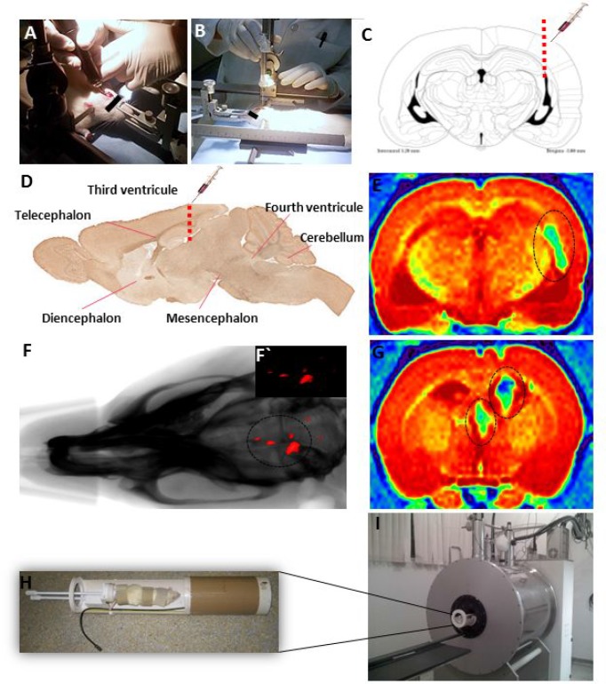
Figure 5.
(A, B) Stereotaxic implantation of EPN cells in brain tumor experimental models. (C) Drawing based on Swanson’s Stereotaxic Atlas Guidelines for the indicated coordinates from Bregma -3.80 mm (region of infusion into EPN cells). (D) Representative sagittal section demonstrating the injection of MION-Rh labeled EPN cells into the third ventricle. (F) Combined fluorescence and X-ray tomography for in vivo detection of EPN cells. F´) Fluorescence image detail. (E, G) MRI monitoring of EPN cells and MION-Rh detection. (H) Radiofrequency bobine of animal positioning. I) MRI equipment for small rodent neuroimaging: 2 Tesla superconducting magnet 85310HR.

