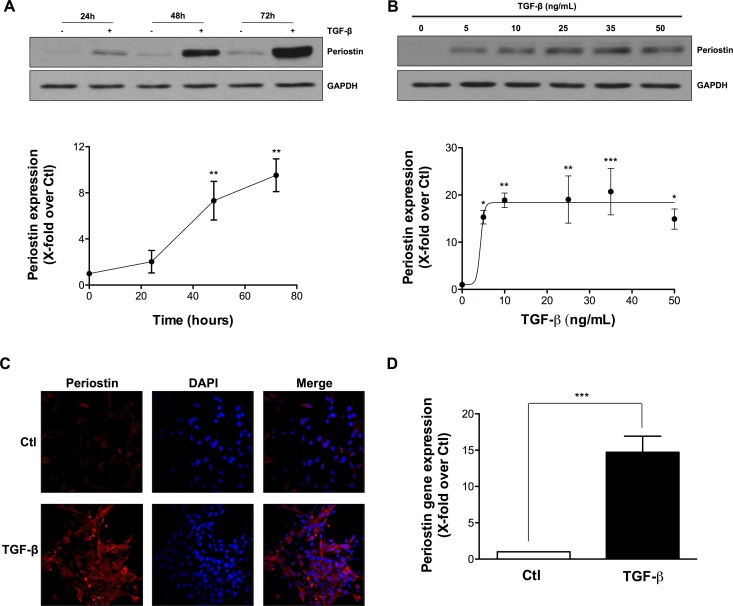
Figure 1. TGF-β induces periostin expression in U-87 MG Cells.
U-87 MG cells were exposed to TGF-β. Western blot analysis demonstrated levels of periostin protein expression after (A) treatment with 10 ng/mL TGF-β for 24, 48 and 72 h or (B) treatment with different concentrations of TGF-β for 48 h. Densitometric analysis is representative of three or more independent experiments. (C) Photomicrographs show the immunostaining of periostin (red) and nuclei (blue) using fluorescence microscopy. (D) The effect of TGF-β on periostin gene expression was evaluated by real-time qPCR. Statistically significant differences were calculated by unpaired Student’s t test (D) and one-way ANOVA followed by Bonferroni’s test (A, B) (*P < 0.05, **P < 0.01, and ***P < 0.001 versus control cells).

