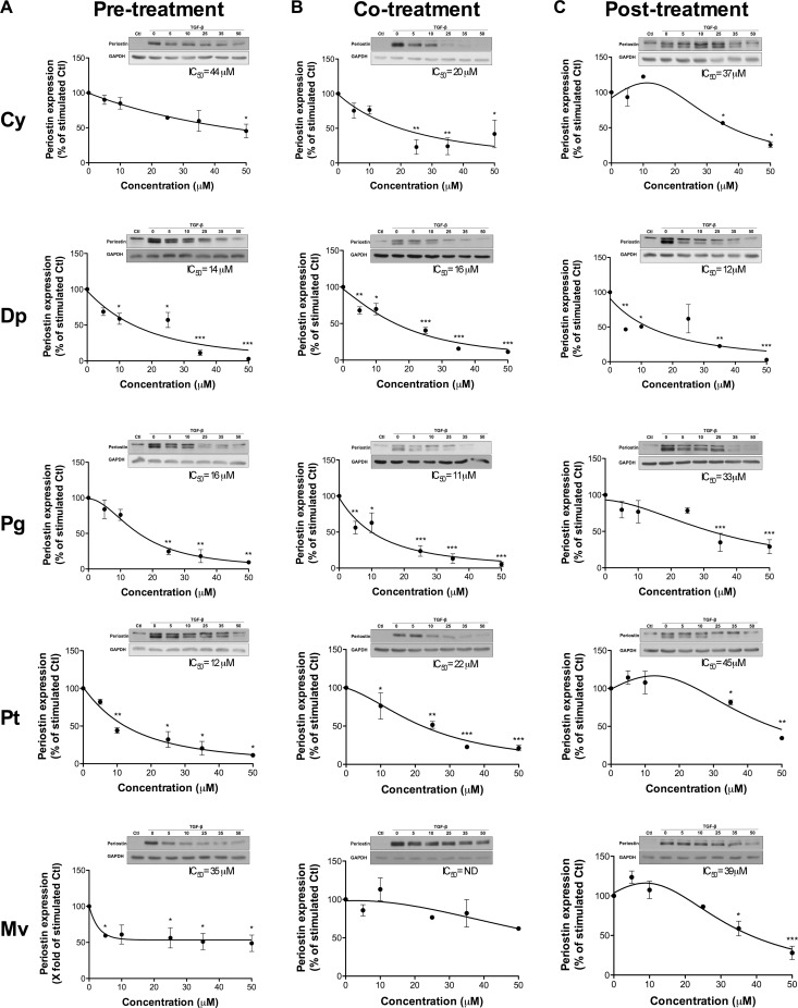
Figure 4. Anthocyanidin effects on the expression of TGF-β-induced periostin.
U-87 MG cells were treated with various concentrations of each anthocyanidin in serum-free medium (A) prior to, (B) along with, or (C) following addition of 10 ng/mL TGF-β. Cells were lysed and the periostin protein levels assessed by immunoblotting. The immunoreactive band intensities were analyzed by densitometry using ImageJ software and expressed as a ratio of levels of periostin to those of the housekeeping GAPDH protein to correct for variations in the amount of proteins loaded. The relative levels of proteins were also normalized to the TGF-β-stimulated condition (value = 100%). Statistically significant differences were calculated by one-way ANOVA followed by Bonferroni’s test (*P < 0.05, **P < 0.01, and ***P < 0.001 versus stimulated control). Data are representative of three or more independent experiments.

