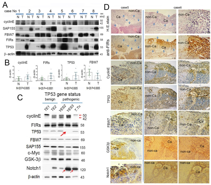
Figure 1. FBW7 was significantly decreased whereas FIRs, cyclin E and TP53 were significantly increased in esophageal cancer tissues indicated by Western blot analysis.
(A) Western blot analysis of cyclin E, SAP155, FBW7, FIRs, and TP53 expression in esophageal cancer tissue (T) and non-cancer tissues (N) without chemoradiation therapy before operation. β-actin was used as internal control. Cyclin E, FIRs, and TP53 were significantly increased in cancer tissues than in non-cancer tissues. On the other hand, FBW7 expression was significantly decreased in cancer tissue. Note, in case 2, TP53 was paradoxically highly expressed in (N) than that of (T). (B) Histogram of protein expression between cancer and non-cancer tissues. The ratio of qRT-PCR results were cyclin E (T/N)=2.0, FIRs (T/N)=2.6, TP53 (T/N)=3.3 and FBW7 (T/N)=0.54. Statistical analysis was performed by t-test. P values < 0.05 were considered significant. (C) Correlation of T/N ration between TP53 and FBW7 was indicated. There was weak negative correlation between TP53 (T/N) and FBW7 (T/N). (D) Cyclin E, FIRs, TP53, FBW7, SAP155, c-Myc, GSK-3β, and Notch1 expression were indicated depending on the TP53 mutational status. β-actin was used as an internal endogenous control. TP53 variation status was benign in HeLa, TE1 and TE2 whereas pathogenic in YES, YES3 and T.Tn. The TP53 was truncated in YES3 cells (thick arrow). Cyclin E was less expressed whereas Notch 1 was increased in YES3 and T.Tn, than those of benign cells (thin arrows). FIR, cyclin E, TP53, FBW7, GSK-3β, Notch1 expression and H&E staining was performed by immunohistochemical staining in esophageal cancer (Ca) and corresponding non-cancer tissues (non-Ca).

