Figure 7.

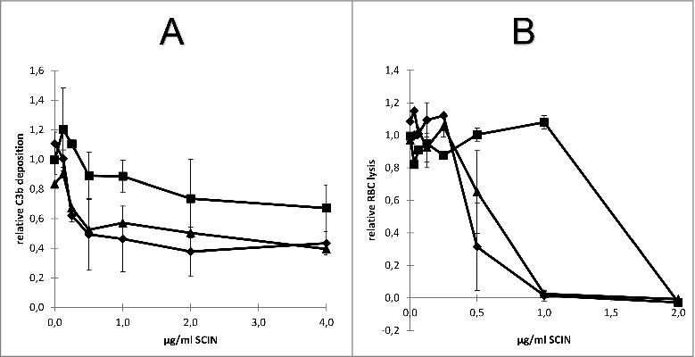
Impact of humAb 6D4 on SCIN activity. (A) C3b deposition on S. aureus Newman ΔspaΔsbi cells upon preincubation of SCIN with humAb 6D4 (■). C3b deposition was monitored by flow cytometry. As a negative control, SCIN was preincubated with buffer (♦), or control IgG (▴). Each data point represents the mean ± standard error (error bars) of 3 independent experiments. (B) Reduced SCIN-mediated protection of rabbit erythrocytes against lysis by complement upon incubation of SCIN with humAb 6D4 (■). Hemolysis was quantified by pelleting of erythrocytes and subsequent measurement of the absorbance of supernatants at 450 nm. As a control, SCIN was preincubated with buffer (♦), or control IgG (▴). Each data point represents the mean ± standard error (error bars) of 2 separate experiments.
