
Figure 6.
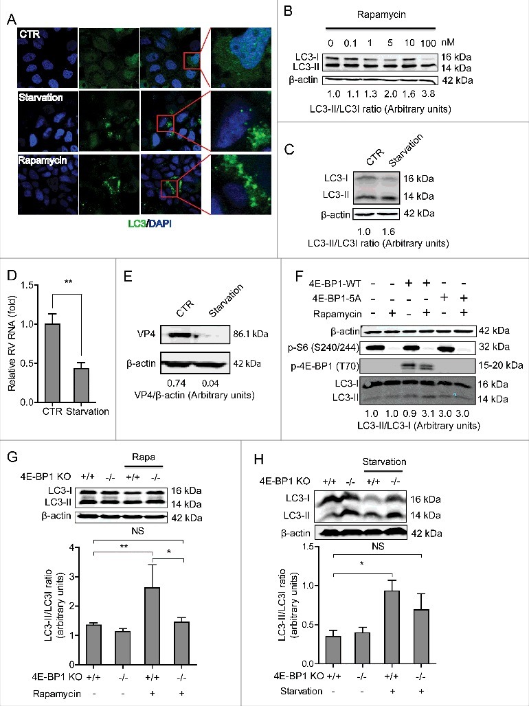
4E-BP1 mediates rapamycin-induced autophagy that inhibits rotavirus infection. (A) Caco2 cells transduced with lentiviral particles carrying a construct of TagGFP2-LC3 driven by the elongation factor-1 promotor were cultured at 37°C for 48 h in DMEM medium containing EBSS medium containing 1mM pepstatin A and E-64-d solution (for starvation) and 10 nM rapamycin. LC3-positive puncta was observed by confocal laser microscopy. (B) LC3-I and LC3-II protein levels were examined by western blot analysis. Protein samples were extracted from Caco2 cells treated with indicated concentrations of rapamycin (48 h). Quantification of the intensity of the immunoreactive bands of both LC3-I and LC3-II was performed using Odyssey V3.0 software. Densitometric analysis of immunoblots of LC3 was expressed as the ratio of LC3-II to LC3-I, and the ratio of LC3II/LC3I was expressed in arbitrary units. (C) Western blot visualized LC3-I and LC3-II protein levels in starvation (EBSS medium containing 1mM pepstatin A and E-64-d solution) treated Caco2 cells. The ratio of LC3II/LC3I was expressed in arbitrary units. (D) Starvation significantly inhibited rotavirus RNA in Caco2 cells (n = 6, mean ± SEM, *P < 0.05, Mann-Whitney test). (E) Starvation dramatically inhibited rotavirus protein VP4 synthesis in Caco2 cells. (F) Rapamycin induced autophagy in 4E-BP1 KO MEFs transfected with 4E-BP1-WT plasmids; while the induction was abolished in 4E-BP1 KO MEFs transfected with 4E-BP1–5A plasmids. (G) Western blot demonstrated that upregulated ratio of LC3-II to LC3-I by rapamycin was abolished in 4E-BP1 knockout (−/−) MEFs (n = 6, mean ± SEM, *P < 0.05, **P < 0.01, Mann-Whitney test). (H) Western blot detected that upregulated ratio of LC3-II to LC3-I by starvation (EBSS medium containing 1mM pepstatin A and E-64-d solution) was abolished in 4E-BP1 knockout (−/−) MEFs (n = 5, mean ± SEM, *P < 0.05, Mann-Whitney test).