
Figure 2.
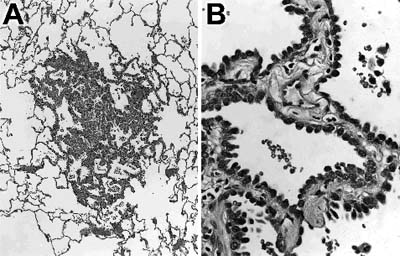
Histologic section of an atypical adenomatous hyperplasia. (A) The lesion shows preservation of normal lung architecture and is surrounded by normal lung parenchyma. (B) Higher magnification shows a single layer of cuboidal cells with cytologic atypia lines septae that are mildly thickened.