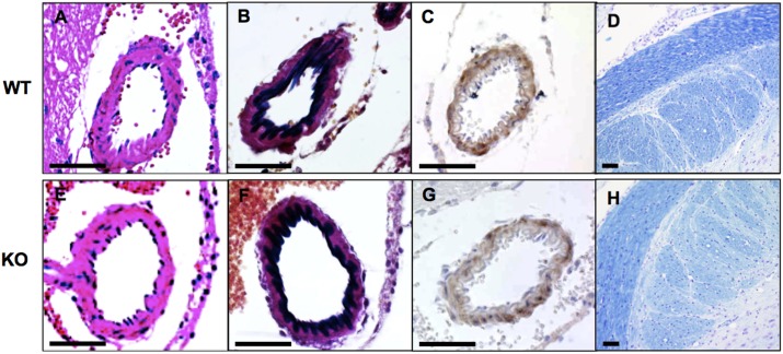
Fig 1. Histology of brain arteries of wild type and HtrA1-/- mice.
Coronal sections of midbrains from 52-week-old wild type (WT) and HtrA1-/- (KO) mice from the 129/B6 background were stained with hematoxylin and eosin (A, E), elastica van Gieson for elastic fibers (B, F), anti-smooth muscle α-actin antibody, a VSMC marker (C, G), and luxol fast blue and cresyl violet for myelin (D, H). No obvious defects were observed in HtrA1-/- brain arteries. Bars = 50 μm.

