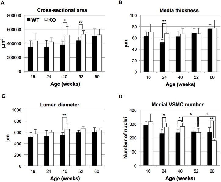
Fig 2. Histomorphometric parameters of aortas isolated from HtrA1-/- mice.
Cross sections from the upper half of the descending aorta were stained with elastica van Gieson (EVG), hematoxylin and eosin (HE), or picrosirius red. The EVG-stained sections were analyzed for A, cross-sectional area; B, media thickness; C, lumen diameter; the HE-stained sections were analyzed for D, medial VSMC number. Bars represent means ± SD (four to 10 mice were used for each group and 3–8 aorta sections per mouse were analyzed. The details are described in S2 File). Statistical significance was determined by Student’s t-test. Asterisks show significance between wild type (WT; black bars) and HtrA1-/- (KO; white bars) mice at the same age (*, p <0.05; **, p <0.01). $ or # show significance (p <0.05) of the difference in medial VSMC numbers between 40- and 52-week-old, or 52- and 60-week-old, HtrA1-/- mice, respectively.

