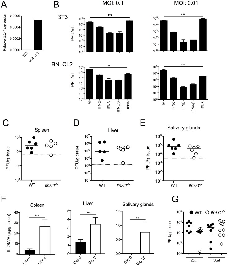
Fig 1. IFNλ can restrict mCMV replication in vitro.
(A) Ifnλr1 expression by 3T3 and BNLCL2 cells was determined by qPCR. (B) 3T3 (top) and BNLCL2 (bottom) cells were incubated with/without 50U/ml IFNα and/or IFNβ, or 50ng/ml IFNλ2 (IL-28A) for 24hrs and infected with mCMV at multiplicities of infection (MOI), as stated in the figure. After 4 days, infectious virions in supernatant were quantified by plaque assay. Statistical significance of PFU in IFNλ2-treated versus control cells is shown. Virus load in spleen (C), liver (D) and salivary glands (E) of WT and Ifnλr1-/- mice was assessed 4 (D&E) and 33 (E) days p.i. (F) IFNλ2/3 protein in spleen (left), liver (middle) and salivary glands (right) was measured at day 0 and 2 days p.i (spleen and liver) or 0 and 26 days p.i (salivary glands). Results are shown as mean + SEM of 3–7 mice/group. (G) WT and Ifnλr1-/- mice were infected (i.n) with mCMV in a volume of 25μl or 50μl and after 4 days, lung infectious viral load was quantified by plaque assay. Statistical significance was assessed using 1-way ANOVA (B) or Mann Whitney-U (C-E, G) or students T-Test (F) and is depicted where appropriate. Panel G represents merged data from two experiments whereas all other data represent at least two biological replicates performed separately.

