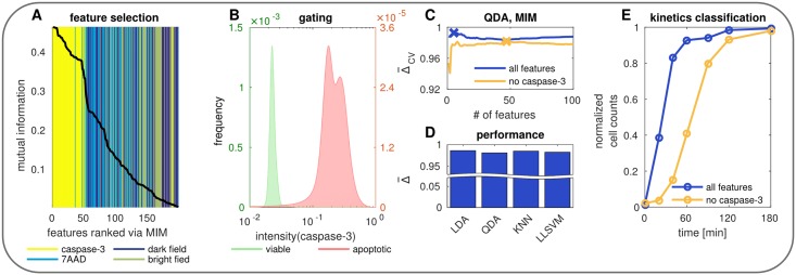
Fig 5. Information content of caspace-3.
(A) Feature selection via MIM. (B) Normalized caspase-3 intensity of viable cells without stimulation (green) and apoptotic (red) cells 180 min after stimulation. (C) Model selection for all features (blue) vs. all features except caspase-3 (yellow). (D) Performance comparison of different classification algorithms trained on all features except caspase-3. (E) Classification of kinetics data using the optimal models from C.

