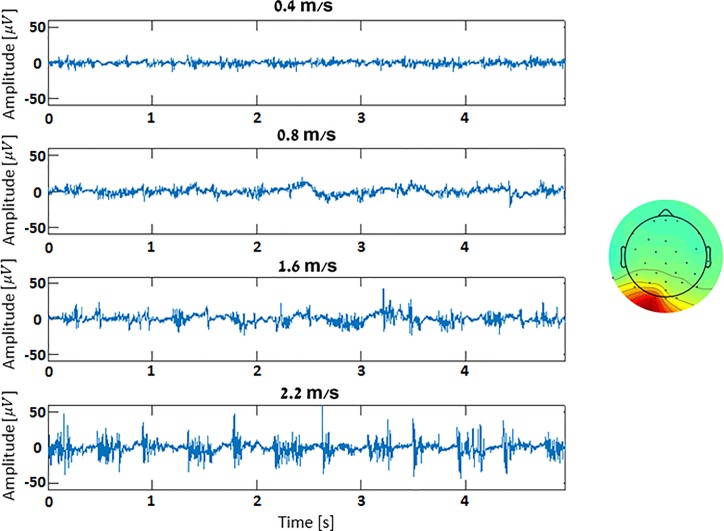
Fig 7. Increased electro-physiological signals during higher speeds.
Presented are 10-second temporal segments of occipital neck muscle components, as separated by the AMICA algorithm (exemplary component map presented on the right), recorded during the various TM speeds of a single participant. Speeds increase from top to bottom, x and y axes depict time and amplitude [V], respectively.

