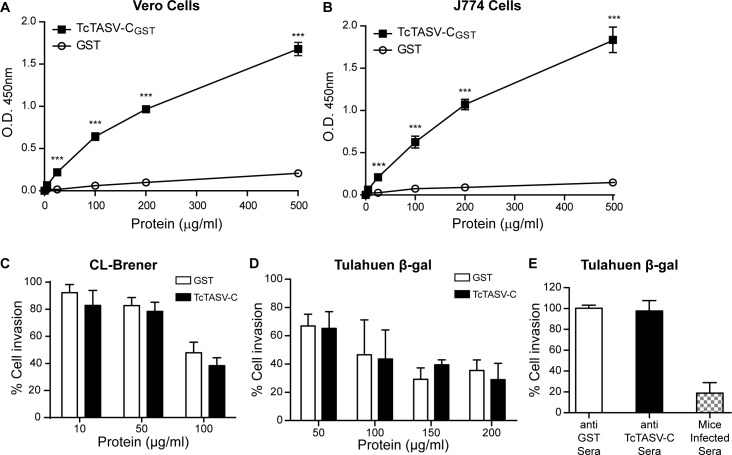
Fig 6. TcTASV-C interacts with mammalian cells but not interfere with T. cruzi cellular infection.
TcTASV-CGST (black squares) or GST (open circles) were incubated with non-phagocytic professional cells (Vero cells) (A) or with professional phagocytic cells (J774) (B). Binding was assessed with a polyclonal anti-TcTASV-CGST sera, in an ELISA-like assay. Values are means ± standard deviation of one assay (run by triplicate) that is representative of 3 independent experiments. (***p< 0.005 vs GST; Student’s t test). (C) Vero cells were pre-incubated with rTcTASV-C for 30 min and then infected with CL-Brener trypomastigotes O.N and washed. After 48 hs cells were fixed and stained with May-Grünwald Giemsa. Infected cells were enumerated by microscopy. (D) Cells were treated as in C, and infected with purified bloodstream trypomastigotes (Tul-β-gal) for 18 hs. Then cells were washed and cultured for 72 h. β-galactosidase activity was measured with CPRG, after cell lysis. (E) Alternatively, purified bloodstream trypomastigotes (Tul-β-gal) were pre-treated with anti TcTASV-C, anti-GST or sera from infected mice for 30 min, before cell infection. Cell infection and measurements were as in D. Data were normalized to untreated control group, considered as 100% of infection.

