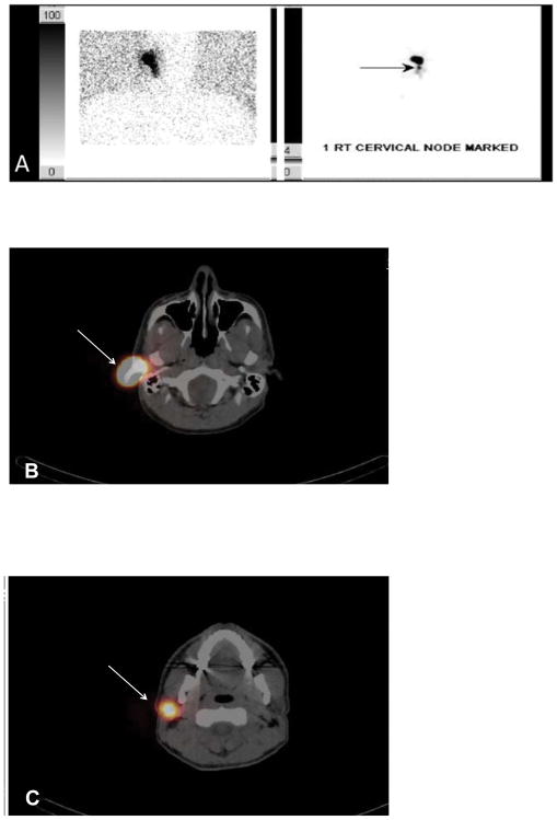
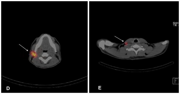
FIG. 1.
(A) Planar lymphoscintigraphy revealed a right ear primary along with one right cervical sentinel lymph node. SPECT/CT demonstrated the primary site (B) and one parotid sentinel lymph node directly inferior to the primary (C) (white arrows). SPECT/CT also demonstrated one node in the right lateral neck level IIB (D) and one in level IV (E) (white arrows)


